
Motor control impairments associated with hip osteoarthritis
Prior to 2009, high quality research and evidence regarding the muscle function changes associated with hip joint disease was lacking. Grimaldi and colleagues published two articles which investigated the association between degenerative hip joint pathology and size of gluteus medius (GMED), gluteus minimus (GMIN), piriformis (PIRI) (Grimaldi, Richardson, Stanton et al., 2009), tensor fascia lata (TFL), and gluteus maximus (GM) muscles (Grimaldi, Richardson, Durbridge et al., 2009).
Hip OA is a considerable problem in modern society and it is becoming more accepted that active rehabilitation plays a vital role in recovery, maintenance and/or prevention of disease progression. "Therapeutic exercise programs designed to improve muscle function around the affected hip will only be maximally effective when we have further information available on both normal muscle function, and changes occurring in association with joint disease" (Grimaldi, Richardson, Stanton, et al., 2009, p. 605).
Purpose
The purpose of these two studies was to examine if there were significant changes in muscle symmetry between patients with no pathology (control group), mild degeneration disease, and advanced hip degenerative disease. Cross sectional areas of each individual muscles were measured using MRI assessment.
Of particular interest for this research, was the functional changes seen in the abductor synergy of the hip. These muscles act to control ground reaction forces during loading and maintain optimal pelvic/hip alignment in both weight bearing and non weight bearing positions. "One of the most consistent findings in subjects with hip dysfunction is the inability to maintain adequate lateral control of the hip and pelvis in single leg stance" (Grimaldi, Richardson, Durbridge, et al., 2009, p. 611).
Results
Changes seen in Mild degenerative disease include:
- There was no significant change in gluteus medius or piriformis present in this group compared to controls.
- In some patients, gluteus medius was larger compared to controls, which may indicate that hypertrophy occurs in the early stages to assist with control of the femoral head.
- The lower gluteus maximus tended to be smaller on the affected side, but the results were not statistically significant.
- There was no change in TFL, in fact, it tended to be larger on the affected side.
Changes seen in Advanced degenerative disease include:
- There is a significant difference in size of gluteus medius and piriformis, both of which were smaller when compared to the control and the mild joint pathology group.
- Gluteus minimus tended to be smaller on the affected size, but the results were not statistically significant.
- Gluteus maximus tended to be smaller on the affected side, mostly in the lower portions of the muscle.
- There was no change in TFL size.
These two studies revealed two key messages.
Hypertrophy/Atrophy associated with joint degeneration is not equal amongst the superficial and deep muscles of the abductor synergy.
Hypertrophy/Atrophy will vary depending on the stage/amount of degeneration.
(Cleland, 2005, p. 249)
Clinical relevance
"Information from this and our previous study together demonstrate that the abductor synergy does not respond homogeneously to joint pathology. While deeper abductor muscle GMED, PIRI, and GMIN demonstrate atrophy in subjects with advanced OA, superficial abductor muscles UGM and TFL appear less affected by underlying pathology" (Grimaldi, Richardson, Stanton, et al., 2009, p. 609).
In clinical assessment, strength testing of abduction often addresses the abductor synergy as a global unit. However, "if both superficial abductor muscles are not significantly affected by pathology, strength changes may possibly reflect weakness in the deeper abductor muscles" (Grimaldi, Richardson, Durbridge, et al., 2009, p.616).
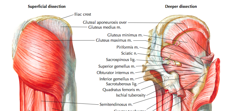
The abductor synergy can be divided into superficial and deep muscles for clinical assessment, based on their attachment.
- Gluteus maximus and tensor fascia lata are both superficial muscles which attach to the iliotibial band.
- Gluteus medius, gluteus minimus, and piriformis are deep muscles which attach to the greater trochanter of the hip.
Therefore, careful assessment of the superficial and deep muscles of the abductor synergy will lead to accurate identification of impairments in muscle function, allowing therapists to create specifically tailored rehabilitation programs.
An overview of the anatomy of the hip and clinical assessment will be the focus of the second part of this blog. Please refer to "Muscle synergies of the hip and pelvis", which will be coming soon....
Sian
References
Cleland, J. (2005). Orthopaedic clinical examination: an evidence-based approach for physical therapists. WB Saunders Co.
Grimaldi, A., Richardson, C., Stanton, W., Durbridge, G., Donnelly, W., & Hides, J. (2009). The association between degenerative hip joint pathology and size of the gluteus medius, gluteus minimus and piriformis muscles. Manual therapy, 14(6), 605-610.
Grimaldi, A., Richardson, C., Durbridge, G., Donnelly, W., Darnell, R., & Hides, J. (2009). The association between degenerative hip joint pathology and size of the gluteus maximus and tensor fascia lata muscles. Manual therapy, 14(6), 611-617.